 EUGENE, Ore. -- (July 22, 2009) -- Applying biological molecules from cell membranes to the surfaces of artificial materials is opening peepholes on the very basics of cell-to-cell interaction.
Two recently published papers by a University of Oregon biophysicist and colleagues suggest that putting lipids and other cell membrane components on manufactured surfaces could lead to new classes of self-assembling materials for use in precision optics, nanotechnology, electronics and pharmaceuticals.
Though the findings are basic, they provide new directions for research to help understand nature at nanotechnological scales where the orientation of minuscule proteins is crucial, said Raghuveer Parthasarathy, who is a member of the UO's Material Science Institute, the Institute of Molecular Biology and the Oregon Nanoscience and Microtechnologies Institute (ONAMI).
The studies, while separate, provide examples of two of the research directions in his lab.
•Controlling interactions between colloidal materials
In the May issue of Soft Matter, a journal of the Royal Society of Chemistry, UO doctoral student Yupeng Kong and Parthasarathy applied biological material -- a thin layer of membrane lipids -- onto to tiny glass spheres about one-millionth of a meter in diamter to closely study colloidal interaction.
Colloids are tiny particles found dispersed in liquids: in milk, paints, many food stuffs, cosmetics and pharmaceuticals. Compared to atoms and molecules colloids are big, and creating artificial colloids with directed properties is a goal in many technologies, especially optics at nanoscales.
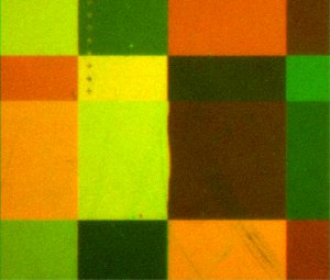
Before applying the biomembrane, the identical negatively charged spheres repelled each other. With the membrane attached, conditions changed dramatically. Suddenly, the like-charged spheres were attracted to each other.
"This was weird," Parthasarathy said. "Like-charged objects aren't
supposed to attract each other. People have seen like-charge attraction
in a few other colloidal systems in the last 10 or 15 years, but still
no one understands it. Here, we've got the first system in which
like-charge attraction can be controlled, simply by the incorporation
of molecules from biological membranes. We can tune attraction or
repulsion over the entire spectrum simply by changing the composition
of the membrane. This is useful both for technological applications,
and for illuminating the fundamental mechanisms behind colloidal
interactions."
The observations were made using an inverted microscopy technique in which the glass spheres were placed in a 655-nanometer diode laser beam, an approach developed in Parthasarathy's lab by former undergraduate biophysics student Greg Tietjen, now a doctoral student at the University of Chicago.
The findings of the National Science Foundation-funded research, he said, suggest that specially tweaked biological membranes applied to artificially produced materials may serve as specialty control knobs that direct materials to do very specific things.
•Controlling molecular orientation from cell membranes:
In a paper appearing online in the Journal of the American Chemical Society (JACS) in early July, Parthasarathy teamed with organic chemists at the University of California, Berkeley, to study how molecules are oriented on their cell membranes to allow for cell-to-cell interactions.
The six-member research team built tiny artificial molecules that mimic brush-like membrane proteins and contain tiny fluorescent probes at the outer end. These miniscule polymers were incorporated into artificial membranes placed on a silicon wafer that acts like a mirror, allowing precise optical measurements of the orientation of the molecule.
Electron microscopy revealed the presence of rigid, rod-like brushy glycoprotein (sugar-containing compounds) -- 30 billionths of a meter long -- similar to natural cell-surface proteins. Interaction between cells occurs when these rods stand up from the membranes, a property whose control remains poorly understood.
The surprise, Parthasarathy said, was that the sugar-laden rods stood up like trees rising in a forest only for particular fluorescent probes, which represented just 2 percent of the molecule's weight.
The big issue that surfaced from the project -- funded by the U.S. Department of Energy, National Science Foundation and the Alfred P. Sloan Foundation -- was that the slightest trepidation of a molecule's structure affects its orientation, he said.
The goal, Parthasarathy said, may be to determine how to control the orientation of the brush-like forest through either chemical or optical measures to, in turn, control cell interaction. Such control of artificially produced molecules, he added, could have huge potential applications in the electronics industry.
Parthasarathy's UO team is now looking at DNA anchored to membranes to compare the findings and see if such on-off switching of the orientation of molecules may be possible.
"There are brush-like proteins at cell surfaces that are really important for such things as cellular interactions within the immune system," Parthasarathy said. "At the surface of every cell is a forest of molecules to induce interactions. These proteins need to rise from the forest. What allows them to stick up or lie down? We've really had a poor idea of what's going on. Knowing the genome and what proteins are there is crucially important, but that information in itself does not tell you anything about the answer to the question."
Co-authors of the JACS study with Parthasarathy are Kamil Godula, David Rabuka, Zsofia Botyanszki and Carolyn R. Bertozzi, all of UC-Berkeley, and Marissa L. Umbel, then an undergraduate student from Indiana University of Pennsylvania who worked in Parthasarathy's UO lab in summer 2008 as part of the UO's National Science Foundation-funded Research Experiences for Undergraduates. Umbel now is studying medical physics at Ohio State University.
