Soft X-ray Speckle Metrology
of Magnetic Multilayer Films
Primary Collaborators:
L.B. Sorensen, M. Pierce, R. Moore,
J.B. Kortright, Materials Sciences Division, LBNL
O.E. Hellwig and E.E. Fullerton, Hitachi
Karine Chesnel, Mark Pfeifer, Advanced Light Source,
LBNL
Josh Turner,
An
important feature of many classes of materials is that the energetic
interactions that determine their thermodynamic and mechanical properties arise
from weak forces that operate on the nanometer length scale. These interactions nonetheless manifest
themselves on a macroscopic scale in ways that lead to unusual and useful
properties. These statements apply as
well to superconductors and ferromagnets as they do to complex fluids and
biological materials. Despite the many
spectacular advances made in developing new microscopy, spectroscopy, and
scattering techniques, a mechanistic understanding of this
microscopic-macroscopic connection has not been achieved in many cases. Part of the reason for this is that most
techniques do not provide simultaneous spatial and dynamical information on key
length and time scales. Diverse
phenomena that involve, for example, thermal activation or exotic phase
separation, can only be partially studied at present because the important
microscopic modes are characterized by nanometer length scales and microsecond
time scales - a regime that is not well-covered by existing experimental
techniques.
In
a growing collaboration, we are developing a technique to cover this
spatio-temporal regime based upon the scattering of transversely coherent beams
of soft x-rays.[1-3] Similar
efforts are underway at other facilities around the world.[4-8]
Conceptually, these techniques constitute a blend of small angle neutron
or x-ray scattering, which probe static density fluctuations on a nanometer
length scale, with dynamic laser light scattering, which probes temporal
fluctuations on a microsecond time scale albeit at a length scale comparable to
the wavelength of visible light. Thus,
the goal is to produce a beam of soft x-rays that have some laser-like
properties, and to utilize this to do dynamic scattering. We utilize the high optical brightness of the
Advanced Light Source in
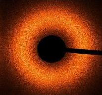
Fig. 1: Speckle-diffraction pattern
of a Pt:Co multilayer, collected at a wavelength of 1.6 nm (very close to the
Co L-edge). Scattering contrast is provided by the huge magneto-optical
variation near the edge. The black stripe and center are the shadow a blocker
that eliminates the direct beam. The pattern is collected in transmission.
Recently,
we have turned our attention to measuring spatial and temporal fluctuations in
magnetic thin films, in collaboration with Jeff Kortright at LBNL and Eric Fullerton
at IBM Almaden Laboratory. By operating
at the relevant 3d transition metal L-absorption edge, it is possible to
achieve marked magnetic contrast.[9] We have
completed extensive measurements of the static speckle patterns produced by
magnetic domain structures of Co:Pt multilayers, an example of which is shown
in Fig. 1 above. This material system
exhibits perpendicular anisotropy and, therefore, is being intensively studied
as a candidate for next-generation recording material. Speckle patterns such as these have been used
to test the notion of microscopic return point memory. Specifically, we have measured the degree to
which the microscopic magnetic domain structure reproduces itself after
traversing major and minor magnetization loops.
We find for this system that the degree of microscopic return point
memory upon traversing a major loop depends critically on the level of
microscopic inhomogeneity of the film.
A new beamline and
scattering apparatus has recently been commissioned and is being used to study
a variety of complex materials and thin film structures.
Acknowledgement:
This work was carried out
in part at the Advanced Light Source at Lawrence Berkeley National Laboratory
which is supported by the U.S. Department of Energy. Financial support from the
USDOE under grant DE-FG06-86ER45275.
References
1. Pierce, M.S., et al., Quasistatic X-Ray Speckle Metrology of Microscopic Magnetic Return-Point Memory. Phys. Rev. Lett., 2003. 90: p. 175502.
2. Price, A.C., et al., Soft X-Ray Dynamic Light Scattering from Smectic A Films. Phys. Rev. Lett., 1999. 82: p. 755.
3. Hu, B., et al., Coherent Soft X-ray Magnetic Scattering. Synch. Rad. News, 2001. 14(2): p. 11-19.
4. Sutton, M., et al., Observation of speckle by diffraction with coherent X-rays. Nature, 1991. 352: p. 608.
5. Cai, Z.H., et al., Observation of X-ray speckle by coherent scattering at grazing incidence. Phys. Rev. Lett., 1994. 73: p. 82.
6. Brauer, S., et al., X-ray intensity fluctuation spectroscopy observation of critical dynamics in Fe3Al. Phys. Rev. Lett., 1995. 74(11): p. 2010-13.
7. Dierker, S.B., et al., X-ray Photon Correlation Spectroscopy Study of Brownian Motion of Gold Colloids in Glycerol. Phys. Rev. Lett., 1995. 75: p. 449.
8. Mochrie, S.G.J., et al., Dynamics of block copolymer micelles revealed by X-ray intensity fluctuation spectroscopy. Phys. Rev. Lett., 1997. 78(7): p. 1275-8.
9. Kortright, J.B. and S.-K. Kim, Resonant magneto-optical properties of Fe near its 2p levels: Measurement and applications. Phys. Rev. B, 2000. 62(18): p. 12216-28.